Normal Oxygen Saturation (‘sats’)
June 26, 2026
Normal oxygen saturation (‘sats’) Oxygen saturation, often referred to as “sats,” measures how much oxygen your red blood cells are carrying. In 2026, with the widespread use o...


The lungs act as the body’s gas exchange system. They process thousands of litres of air every day to deliver vital oxygen to the bloodstream and remove waste gases.
An adult at rest breathes roughly 12 to 20 times per minute, which adds up to around 17,000 to 30,000 breaths every single day. With each normal quiet breath, you inhale about 0.5 litres of air.
Over a 24-hour period, the lungs continuously move more than 11,000 litres of air through their passages.
An adult’s total blood volume passes through the lungs once every minute, meaning the entire circulation is constantly exposed to the air sacs for filtration and gas refresh.
Although they are light and filled with air, if you unfolded all the microscopic air sacs inside the lungs, their total surface area would cover a space roughly the size of a tennis court (about 70 to 100 square metres).
Their main roles are to:
Extract oxygen – from the air and transfer it directly into the bloodstream to power every cell in the body
Excrete carbon dioxide – a major waste product of metabolism, removing it from the blood and exhaling it into the atmosphere
Maintain stable acid-base balance – by adjusting how fast carbon dioxide is breathed out to keep blood pH within a safe range
Protect the body – by filtering out dust, pollutants, and airborne pathogens using a built-in defence system
The lungs are part of the respiratory system.
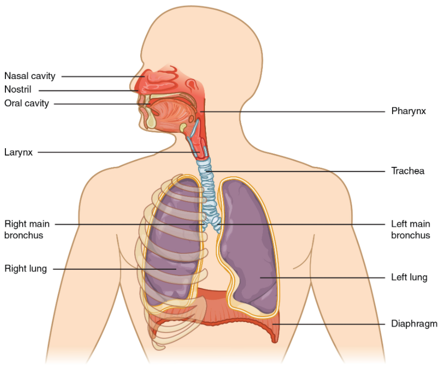 Respiratory System
Location and Size
The lungs are paired, cone-shaped organs that occupy most of the chest cavity (thoracic cavity), sitting on either side of the heart.
Together, they weigh roughly 800 to 1,000 grams, with the right lung typically being slightly heavier than the left.
They sit high up in the chest, protected by the rib cage, extending from just above the collarbones down to the diaphragm muscle at the lower edge of the ribs.
Important note: Sharp or sudden pain when taking a deep breath is rarely coming from the lung tissue itself, as the lungs lack pain receptors. This type of pain usually comes from the surrounding chest wall muscles, ribs, or the sensitive lining around the lungs (the pleura).
The left lung is slightly smaller than the right lung, featuring an indent called the cardiac notch to make room for the heart, which tilts to the left.
Each lung is protected by:
The rib cage – twelve pairs of elastic bones providing a heavy-duty physical shield
The pleura – a double-layered membrane. The outer layer lines the chest wall, while the inner layer wraps the lung tissue
Pleural fluid – a thin layer of lubricant between the pleural membranes that prevents friction, allowing the lungs to expand smoothly
Internal Anatomy
Lobes: The right lung is divided into three sections (superior, middle, and inferior lobes) by deep grooves called fissures. The smaller left lung is divided into just two sections (superior and inferior lobes).
Trachea (Windpipe): The main rigid tube that carries air down from the throat, reinforced by tough C-shaped rings of cartilage to keep it from collapsing.
Bronchi: The trachea splits into two main branches—the left and right primary bronchi—which enter each respective lung.
Bronchioles: Tiny, microscopic branching tubes that split off the bronchi, narrowing further and further down into the deep tissue.
Hilum: The central entry and exit point on the inside edge of each lung, where the main bronchi, pulmonary arteries (bringing oxygen-poor blood in), pulmonary veins (carrying oxygen-rich blood out), and nerves pass through.
Each lung contains roughly 150 to 200 million microscopic air sacs called alveoli, clustered at the very ends of the branching bronchioles.
Every alveolus has two core parts:
Air Sac: A tiny, hollow pocket that fills with fresh air during inhalation. Its wall is incredibly thin—only one cell thick.
Capillary Network: A dense web of microscopic blood vessels wrapping tightly around the outside of the air sac. The capillary wall is also just one cell thick, creating an ultra-thin barrier between air and blood.
Step-by-step process
Air enters the respiratory tract via the nose or mouth, travelling down the trachea and through the branching bronchi.
It flows into the microscopic bronchioles and fills the millions of tiny alveoli sacs.
Oxygen moves passively through the micro-thin walls of the alveoli and capillaries into the bloodstream, binding to red cells.
Simultaneously, carbon dioxide waste moves out of the blood into the alveoli, to be carried up the airways and expelled from the body when you exhale.
Beyond exchanging oxygen and carbon dioxide, the lungs perform several other critical jobs:
Blood pressure regulation: The lining of the lung capillaries produces Angiotensin-Converting Enzyme (ACE), a vital component of the system that regulates systemic blood pressure and fluid balance.
The mucociliary escalator: The airways are lined with a sticky mucus layer and microscopic, hair-like structures called cilia. The cilia beat upward in synchronized waves to sweep dust, bacteria, and debris out of the lungs toward the throat to be swallowed or coughed away.
Blood filtration: The lung capillaries act as a safety filter for the entire circulation, trapping and dissolving tiny blood clots or air bubbles before they can travel to vital organs like the brain or heart.
Phonation: The lungs provide the continuous airflow required to vibrate the vocal cords in the larynx, allowing for speech, singing, and vocal communication.
Respiratory Tract Infections (RTIs)
These are infections caused by viruses, bacteria, or fungi that can affect any part of the breathing system.
Most common colds affect the upper tract (nose and throat).
If an infection travels deep into the tissue, it can cause pneumonia or bronchitis.
Pneumonia causes the delicate alveoli sacs to fill with pus or fluid, making it difficult for oxygen to cross into the blood, resulting in fever, cough, and shortness of breath.
Asthma
A chronic condition characterized by hyper-reactive, inflamed airways that narrow suddenly in response to triggers like dust, pollen, cold air, or exercise.
During an attack, the smooth muscles around the bronchioles tighten up and secrete excess mucus.
This narrows the passages significantly, causing wheezing, chest tightness, and difficulty breathing.
Chronic Obstructive Pulmonary Disease (COPD)
A progressive, long-term condition that causes persistent breathing difficulties due to damage to the airways and air sacs.
Affects hundreds of millions of people globally, with long-term tobacco smoking being the leading cause.
It includes emphysema (where the delicate walls of the alveoli are permanently destroyed, reducing surface area) and chronic bronchitis (long-term inflammation and narrowing of the airways).
Pulmonary Embolism (PE)
A rapid, dangerous blockage in one of the pulmonary arteries inside the lungs.
Usually occurs when a blood clot forms in the deep veins of the legs (Deep Vein Thrombosis, DVT) and travels up through the heart into the lung circulation.
Requires emergency medical treatment, as it blocks blood flow to the gas exchange machinery and strains the heart.
Doctors use a combination of simple checks to evaluate lung health:
Spirometry: The gold-standard lung function test. You take a deep breath and blow out as hard and fast as possible into a machine to measure the total volume of air your lungs can hold and how quickly you can empty them.
Pulse oximetry: A quick, painless clip placed on a finger that uses light waves to measure the oxygen saturation percentage in your blood (normal levels are typically 95% or higher).
Chest x-ray or CT scan: Visual imaging techniques used to look at the structural health of the lungs, checking for signs of infection, fluid, scarring, or masses.
Arterial Blood Gas (ABG): A direct blood sample taken from an artery (usually in the wrist) to measure the exact levels of oxygen, carbon dioxide, and blood acidity in real time.
You can protect your lungs with these simple steps:
Avoid all smoking and vaping: Inhaling tobacco smoke or vaporized chemicals paralyzes the cleansing cilia, triggers chronic inflammation, and permanently destroys the delicate alveoli walls.
Exercise regularly: Cardiovascular activities like brisk walking, running, swimming, or cycling push the lungs to work harder. While it doesn’t change lung size, it trains the heart and muscles to use oxygen more efficiently, reducing overall breathlessness.
Minimise exposure to pollutants: Avoid exercising near heavy traffic or on high-pollution days. Wear appropriate protective masks if working with dust, chemicals, wood shavings, or stone particles.
Practice deep breathing: Take time to practice slow, deep diaphragmatic breathing to ensure the lower sections of the lungs are fully expanding and clearing out old air.
Normal oxygen saturation (‘sats’) Oxygen saturation, often referred to as “sats,” measures how much oxygen your red blood cells are carrying. In 2026, with the widespread use o...
Heart Attack vs. Stroke : Key Differences and Symptoms It is a common question: Is a stroke a type of heart attack? The short answer is no. Whilst both are medical emergencies caused by blood flow iss...
Clopidogrel – a tablet to prevent blood clots Clopidogrel is a tablet for preventing blood clots. What is clopidogrel? Clopidogrel is a commonly used antiplatelet medication prescribed to reduce...
A normal blood cholesterol level Total cholesterol (chol) = 3-5 mmol/L – overall amount of cholesterol in your blood HDL (‘good’) cholesterol. Target level = >1 mmol/L – may...

