1. Definition: The lungs are a pair of pinkish-gray spongy, cone-shaped organs in the chest cavity.
Lung Weight and Size
In terms of size, they are roughly 24 cm (9.4 inches) tall during normal breathing.
Primary Function: When you inhale (breathe in), air enters your lungs, and from that air moves to your blood. At the same time, carbon dioxide, a waste gas, moves from your blood to the lungs and is exhaled (breathed out). This process, called gas exchange, is essential to life.
2. Lung Lobes
Fissures: The lungs are divided into lobes by fissures. The right lung has both an oblique and a horizontal fissure, while the left lung has only an oblique fissure.

Upper and Lower Lobes
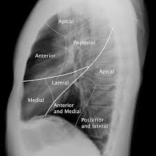 Lateral chest x-ray of R lung
Note. Most of the posterior (back) of the lung is the the lower lobe, and most of the anterior (front) is the upper lobe.
Respiratory System
The lungs are part of the respiratory system: comprising the upper (larynx and above) and the lower (trachea and below) parts.
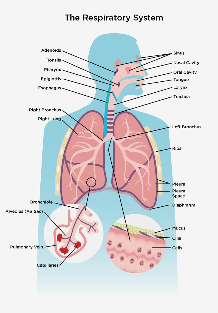
3. Bronchi: The trachea divides into the right and left primary bronchi, each entering a lung. These bronchi further divide into secondary (lobar) bronchi, then tertiary (segmental) bronchi, and continue to branch into smaller bronchioles.
4. Bronchioles: Bronchioles are smaller airways that lack cartilage in their walls. They further divide into terminal bronchioles, leading to respiratory bronchioles, and then alveoli.
5. Alveoli: Alveoli are small air sacs located at the end of the respiratory bronchioles and alveolar ducts. They are the primary sites for gas exchange between the lungs and the bloodstream. There are 300 to 500 million across two lungs.
6. Blood supply
7. Lung Hilum: The hilum of the lung is the area where structures such as the bronchi, pulmonary arteries and veins, and nerves enter and exit the lung.
8. Pleura: The pleura is a double-layered membrane surrounding each lung. The visceral pleura adheres to the lung surface, while the parietal pleura lines the thoracic cavity. The pleural space between these layers contains fluid that facilitates smooth movement during breathing.
9. Diaphragm: The diaphragm is a dome-shaped muscle, that separates the chest cavity from the abdominal cavity, and is the primary muscle used for breathing.
It contracts during inhalation, increasing the thoracic cavity volume and drawing air into the lungs.
10. Lymphatic Drainage: The lungs have a lymphatic network that helps remove foreign particles and excess fluid. Lymphatic vessels are found in the lung parenchyma and along the bronchi and blood vessels.
