1. Definition: The liver is a large dark reddish-brown, wedge-shaped organ that is approximately the size and shape of a rugby ball.
The liver is the largest solid organ in the body, weighing some 1.8 kg in men and 1.3 kg in women. This is 2% of the body weight. It is located in the upper right-hand portion of the abdomen, under the diaphragm and sits above the stomach.
If you place your right hand over the ribs on the lower right side of the front of the chest, it will just about cover the area of your liver.
The liver filters about 1.7 litres of blood per minute. It contains 300 billion specialised cells – yes, alot.
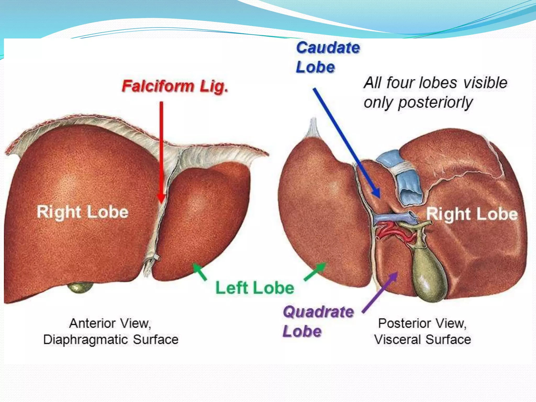
2. Liver Lobes: The liver is divided into four lobes: right, left, quadrate, and caudate. The right lobe is larger and further divided into segments based on the Couinaud classification.
3. Couinaud Classification: The liver is functionally divided into eight segments based on blood supply and drainage. Each segment has its own hepatic arterial and portal venous supply and hepatic venous drainage.
Segmental Autonomy. Each liver segment functions independently with its own vascular inflow, outflow, and biliary drainage. This allows for segmental liver resections while preserving liver function.
4. Liver functions: The liver is the factory of the body, and has over 500 functions. Here are some of the important ones.
5. Blood Supply: The liver is unique in that it receives a dual blood supply: 75% from the hepatic portal vein (deoxygentated blood rich in nutrients) and 25% from the hepatic artery (oxygenated blood). This is crucial for liver function and regeneration.
6. Venous drainage – Hepatic Veins: There are three main hepatic veins (right, middle, and left) that drain into the inferior vena cava (IVC). Understanding their anatomy is crucial for liver resections and IVC filter placement.
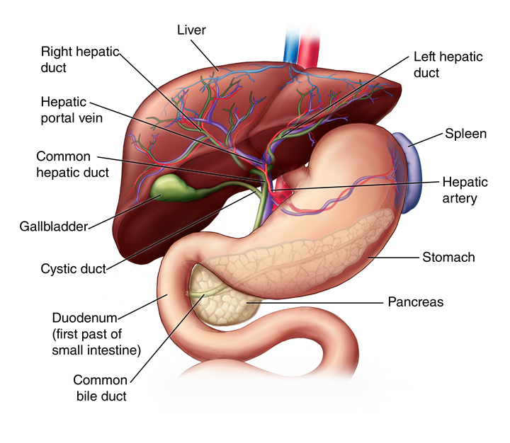
7. Liver Hilum: The liver hilum (porta hepatis) is where the portal vein, hepatic artery, and bile ducts enter or exit the liver. It’s a key area for surgical dissection in liver resections.
8. Bile Production: The liver continuously produces bile, a fluid essential for breaking down fats and helping with the digestive process.
Biliary Drainage: Bile ducts follow the arterial supply. The right and left hepatic ducts join to form the common hepatic duct, which then joins the cystic duct to form the common bile duct.
9. Liver Ligaments: The liver is supported by several ligaments (falciform, coronary, and triangular ligaments) that attach it to the diaphragm and abdominal wall. These are important landmarks during surgery.
10. Liver Regeneration: The liver has an incredible ability to regenerate. If up to 75% of it is removed, it can grow back to its full size in about 6 to 8 weeks.
The ancient Greeks may have somehow known this fact, as in Greek mythology, the gods punished Prometheus for giving humans fire by chaining him to a rock, where a vulture would peck out his liver. Each night his liver would regenerate and the agony would repeat the next day.
The liver is the primary site for blood cell production in the foetus, until the bone marrow fully takes over later in development.
The bulk of the liver is underneath the chest wall, but its inferior edge may occasionally be palpated just below the costal margin. This is mainly in some slim women, on deep inspiration. For most people, the liver is not paplable by a doctor.
Liver anatony (Bazira, 2023)
Liver physiology (Kalra, 2023)
