In this article we describe the size, anatomy and function of the kidney.
1. Location: Most people have two kidneys – they lie under the ribs and above the waist (at the back), one on either side of the body. 1 in 1000 people are born with one kidney and come to no harm. The single (or other if you have one removed) kidney grows (hypertrophies), and the remaining kidney continues to sustain the body.
They are higher up than most people think. The kidneys are protected from injury by a large layer of fat, along with your lower ribs and back muscles.
 This diagram shows the location of the kidneys. The right kidney is slightly lower (and smaller), pushed down by the liver.
The kidneys are retroperitoneal reddish-brown organs located on either side of the spine, between the 12th thoracic vertebra (T12) and the 3rd lumbar vertebra (L3).
Size
In adults, normal kidneys are 10-14 cm long in men and 9-13 cm long in women (12 cm is a good average number), 3-5 cm wide, 3 cm in antero-posterior thickness.
They are are bean-shaped, and about the size of the palm and weigh 150-200 g.
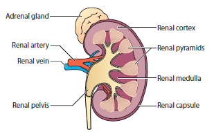
2. Structure: Each kidney is composed of an outer cortex, a middle medulla, and an inner renal pelvis. The renal cortex contains nephrons, the functional units of the kidney.
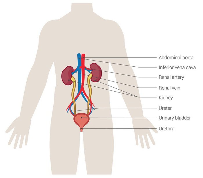
The kidneys are part of the urinary tract.
 Urinary Tract
3. Functions
Note. Two simple tests can detect most cases of kidney disease – a blood test (creatinine/glomerular filtration rate (GFR)) and a urine test (urine Albumin-Creatinine Ratio, ACR).
3. Blood Supply: The kidneys receive blood via the renal arteries, which branch off the aorta. Venous drainage occurs through the renal veins into the inferior vena cava.
Even though the kidney only accounts for 0.5% of the body’s weight on average, it receives more blood (20-25% of the cardiac output) than all other organs except the liver.
4. Nephron: A nephron consists of a glomerulus (where filtration occurs) and a renal tubule (where reabsorption and secretion occur). Each kidney contains approximately 1 million nephrons.
In other words, each kidney has 1 million tiny filters. It s not one big filter.
 Kidney and glomerulus as part of urinary tract
5. Renal Hilum: This is the entry and exit point for the renal artery, vein, and ureter. It’s an important landmark during surgical procedures.
6. Renal Capsule: The kidney is surrounded by a tough fibrous capsule that helps protect the organ and maintain its shape.
7. Collecting System: The renal pelvis collects urine from the calyces and channels it into the ureter, which then transports it to the bladder.
8. Microscopic Structure: The nephron’s glomerulus filters blood, while the proximal and distal convoluted tubules (DCT and PCT) reabsorb most nutrients and water. The loop of Henle regulates water and electrolyte balance.
9. Juxtaglomerular Apparatus (JGA): Located near the glomerulus, the JGA regulates blood pressure and electrolyte balance by controlling renin release.
10. Innervation: The kidneys are innervated by the renal plexus, which includes sympathetic and parasympathetic fibers that regulate renal blood flow and function.
Internally, the kidneys have an intricate and unique structure. The kidney itself can be divided into:
The cortex extends into the medulla, dividing it into triangular shapes – these are known as renal pyramids (10-14 of those) [“nothing to do with the Pyramids of Egypt”. Ed]. The apex of a renal pyramid is called a renal papilla.

Each renal papilla is composed of structures called minor calyces (singular = ‘calyx’; means a cup-like cavity), which collect urine from the pyramids.
Several minor calyces merge to form a major calyx (2-3 of these). Urine passes through the major calyces which unite to form the renal pelvis, a flattened and funnel-shaped structure.
From the renal pelvis, urine drains into the ureter, which transports it to the bladder for storage.
Note 1. A ‘hilum’ is a ‘small thing’ in Latin; and in biology it has come to mean a depression or fissure where structures such as blood vessels and nerves enter an organ
Note 2. ‘Pelvis’ means a ‘basin’ in Latin, from the Greek word pelex (‘helmet’) and pelike (‘goblet’ or ‘bowl’).
Review article: anatomy of kidneys (Soriano, 2023)
