1. Heart Structure: The heart is a muscular organ with a quadrangular pyramid shape. The heart wall consists of three layers: endocardium, myocardium (most of muscle), and epicardium.

It’s roughly the size of a fist and weighs between 200 and 425 grams (7 to 15 ounces), with averages differing between sexes. An adult male heart typically weighs around 315 grams, while an adult female heart averages about 265 grams.
2. Heart Function: The heart’s main function is to pump oxygenated blood to the body’s tissues and organs, removing waste products. It works tirelessly, even during deep sleep, to maintain vital functions.
3. Chambers: The heart has four chambers: two atria (upper chambers) and two ventricles (lower chambers). The atria receive blood, while the ventricles pump blood out of the heart.
4. Valves: The heart has four valves that ensure blood flows in one direction: tricuspid, pulmonary, mitral, and aortic valves. These valves open and close to regulate blood flow.
5. Blood Flow: The heart receives deoxygenated blood through the superior and inferior vena cavae into the right atrium, and oxygenated blood enters the left atrium through the pulmonary veins.
6. Heartbeat: The heart beats around 100,000 times per day, pumping approximately 9000 litres (2,000 gallons) of blood daily. The heartbeat is controlled by the sinoatrial node (SAN), the heart’s natural pacemaker.
7. Electrical System: The heart has its own electrical system, which regulates the heartbeat. Specialised cells called pacemaker cells (in the sino-atrial node, SAN), generate electrical impulses that cause the heart to contract and relax in a regular rhythm.

8. Blood Pressure: The heart creates significant pressure to pump blood throughout the body, equivalent to squirting blood up to 30 feet.
9. Heart Development: The heart is the first organ to form in a foetus, developing early to circulate blood and oxygen to the growing embryo.
10. Heartbeat Sounds: The heartbeat sounds, often described as ‘lub-dub’, are created by the opening and closing of the heart valves as blood flows through its chambers.
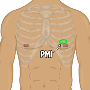
It can be felt during a physical examination. This helps doctors assess the heart’s position, size, and function.
